
Key Takeaways:
- The chronic anterior uveitis associated with juvenile idiopathic arthritis is usually asymptomatic, and the affected eye typically looks entirely normal — white sclera, normal pupil, no redness, no light sensitivity, and no complaint from the child.
- In a population-based Nordic cohort that followed 434 children for eighteen years from arthritis onset, uveitis developed in 96 of them (22.1 percent), and ocular complications occurred in 38.8 percent of the children who developed it.
- American College of Rheumatology and Arthritis Foundation guidance sets the screening interval by risk category rather than by symptoms: a child who is ANA-positive, whose arthritis began at or before age six, and whose disease has lasted four years or less is examined at the slit lamp every three months.
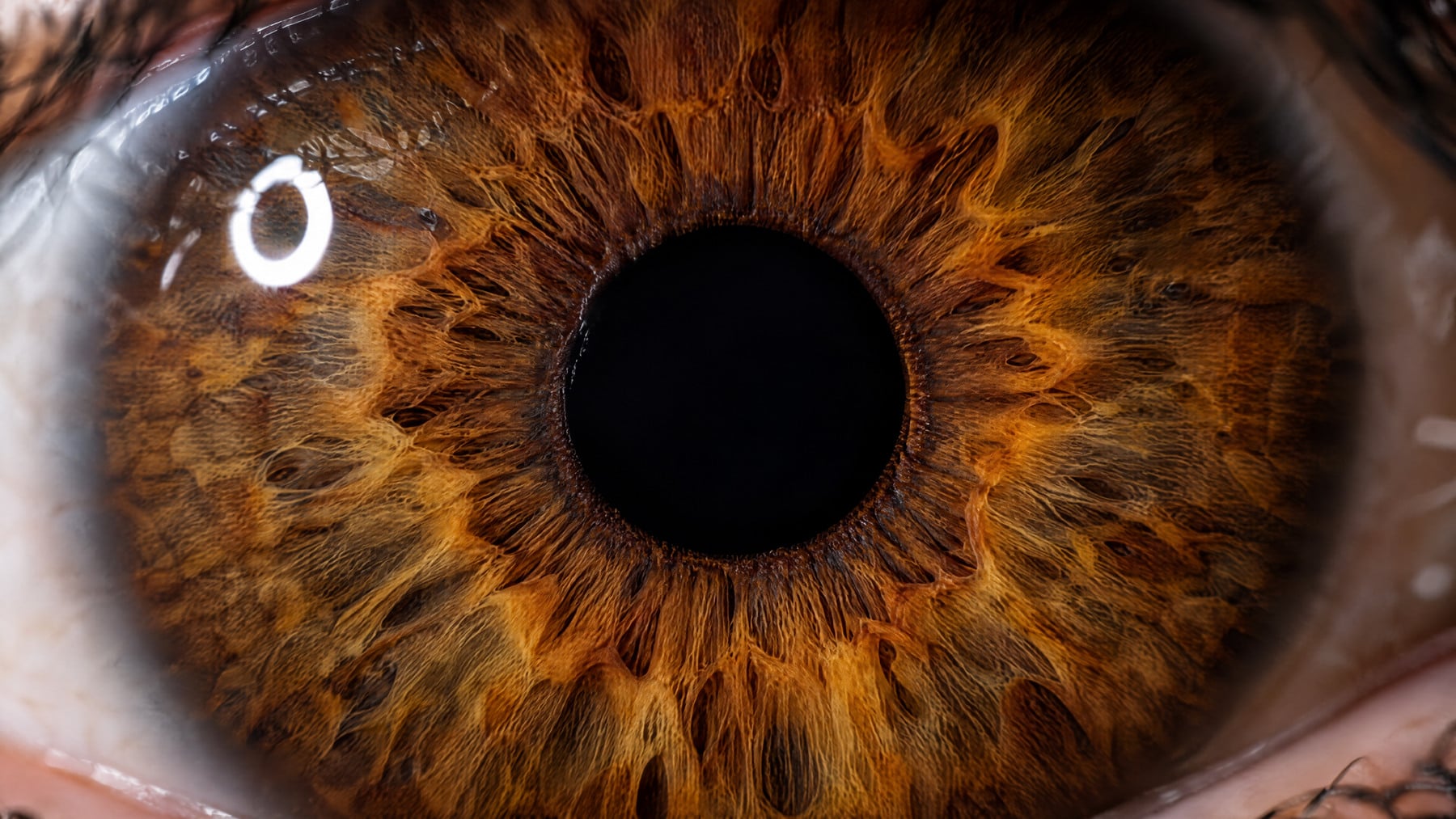
With the school year underway, many children have just passed a school vision screening, and for a child with juvenile idiopathic arthritis that result says nothing about the complication that deserves the most attention. It is not in a joint at all. Juvenile arthritis uveitis — chronic anterior uveitis arising in a child with juvenile idiopathic arthritis — is the most common manifestation of the disease outside the joints, and its defining clinical feature is that it announces absolutely nothing. The process is driven predominantly by CD4-positive T cells in the anterior chamber, and it can run for months while the eye looks entirely ordinary from the outside. White sclera. Normal pupil. No tearing, no photophobia, no rubbing, no complaint. Set that against the acute anterior uveitis of HLA-B27-associated disease, which arrives as a red, aching, light-intolerant eye and sends an adult to an ophthalmologist inside of a day. There the pain is the alarm system, and it works. In the juvenile form, the alarm was simply never wired.
What accumulates behind that quiet exterior is structural rather than symptomatic. Inflammatory cells and leaked protein in the anterior chamber — graded at the slit lamp as cells and flare — settle at the pupillary margin, and fibrinous adhesions bridge the posterior iris to the anterior lens capsule as posterior synechiae, tethering the pupil so it no longer dilates in a round shape. Calcium precipitates in a horizontal band across the exposed cornea as band keratopathy. The lens opacifies, driven both by the inflammation itself and by the topical glucocorticoids used to suppress it. Intraocular pressure climbs — from trabecular meshwork inflammation, from a steroid response, or from synechiae obstructing aqueous outflow — and glaucoma follows. The population-based Nordic cohort, which enrolled children whose arthritis began between 1997 and 2000 and followed them for eighteen years, found uveitis in 96 of 434 children, or 22.1 percent, with ocular complications in 38.8 percent of those affected. Children whose uveitis appeared soon after the arthritis began carried the higher complication risk. Those figures are best read as the case for surveillance, not as a forecast for any individual child.
How often is juvenile arthritis uveitis screened for?
Because symptoms cannot be trusted, the calendar does the work. Risk is not evenly distributed across juvenile idiopathic arthritis; it concentrates in the oligoarticular and rheumatoid-factor-negative polyarticular categories, in children who are antinuclear antibody positive, in those whose arthritis began in early childhood, and in the first several years after onset, when most uveitis appears. American College of Rheumatology and Arthritis Foundation guidance converts those variables into an interval rather than a symptom trigger. A child who is ANA-positive, whose arthritis began at or before age six, and whose disease has been present for four years or less is examined at the slit lamp every three months. Lower-risk categories are seen less often, and intervals lengthen as disease duration grows and the highest-risk window closes. Uveitis occasionally precedes the arthritis entirely, which is part of why an unexplained finding on a child’s eye examination earns a rheumatologic look. The reassuring part is genuine: this is one of the few serious complications in pediatric rheumatology where a kept appointment does most of the protective work. Caught before synechiae and cataract, it responds to treatment — topical glucocorticoids to gain control first, with methotrexate and the monoclonal tumor necrosis factor inhibitors adalimumab and infliximab when systemic therapy is warranted. The appointment a child does not feel they need is precisely the one that matters.
References:
- Clarke, S. L., Sen, E. S., & Ramanan, A. V. (2016). Juvenile idiopathic arthritis-associated uveitis. Pediatric Rheumatology Online Journal, 14(1), 27.
- Rypdal, V., Glerup, M., Songstad, N. T., Bertelsen, G., Christoffersen, T., Arnstad, E. D., et al. (2021). Uveitis in juvenile idiopathic arthritis: 18-year outcome in the population-based Nordic cohort study. Ophthalmology, 128(4), 598-608.
- Angeles-Han, S. T., Ringold, S., Beukelman, T., Lovell, D., Cuello, C. A., Becker, M. L., et al. (2019). 2019 American College of Rheumatology/Arthritis Foundation guideline for the screening, monitoring, and treatment of juvenile idiopathic arthritis-associated uveitis. Arthritis Care & Research, 71(6), 703-716.
Comments are closed